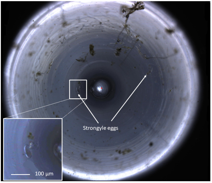
Figure 2.
A zoomed-out view of an equine faecal egg count imaged using the FECPAKG2 Micro-I. The circular image demonstrates a 3 mm field of view at the top of the meniscus produced in the FECPAKG2 Micro-I cassette. Helminth eggs in a flotation solution accumulate to the centre of the well, following the shape of the meniscus. The central circle represents the tip of the light source within the cassette. Equine strongyle eggs are highlighted with white arrows. One of two images produced for each G2 cassette, with each helminth egg seen over the combined two images representing 45 epg, calculated from the dilution factor and the volume of the FECPAKG2 cassette. These images are visible only to the technicians who identify the eggs, and they are large file sizes so they can be zoomed in without loss of clarity in order for accurate identification of helminth eggs.

