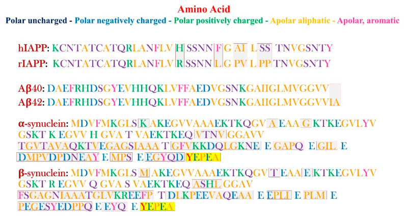
Figure 1.
Schematic representation of the primary sequences of hIAPP, rIAPP, Aβ(1-40) and Aβ(1-42), α and β-synuclein (respectively, from the top to the bottom). The different amino acid residues are pictured in different colors, according to the legend (on the top right of the figure). The differences between the h- and r-IAPP, Aβ(1-40) and Aβ(1-42), α and β-synuclein, respectively, are highlighted by red boxes for the first two pairs, and by red boxes for the last couple of proteins.

