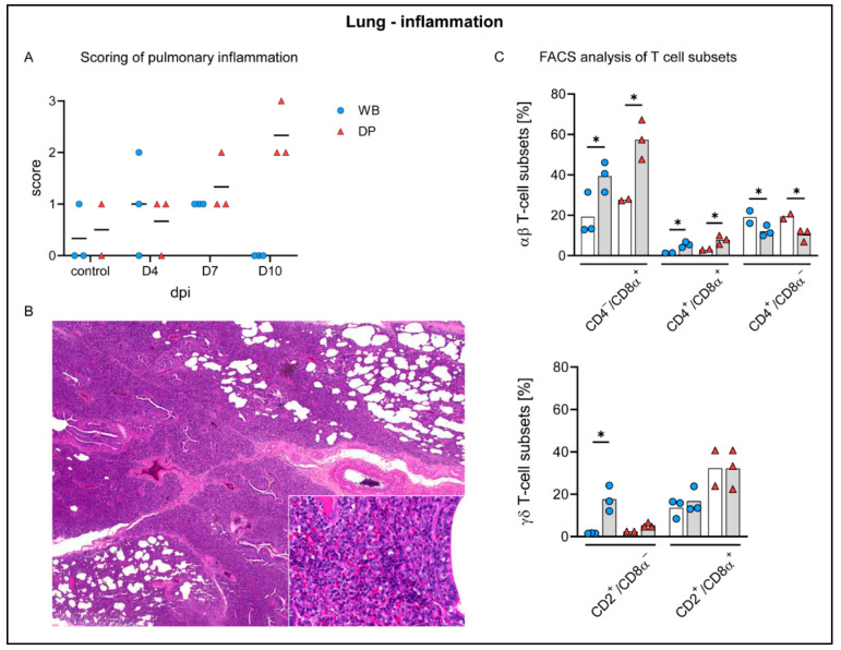
Figure 7.
Pulmonary inflammation in wild boar and domestic pigs. (A) Microscopical scoring of pulmonary inflammation. (B) Section of lung tissue from a domestic pig showing severe interstitial pneumonia with mainly infiltrating lymphocytes and histiocytes (inset) at day 10 pi. (C) Frequency of αβ and γδ T-cell subsets in the lung of control animals (white bars) and wild boar and domestic pigs at 10 dpi (grey bars). WB = wild boar, DP = domestic pig, * p < 0.05, median as horizontal line (A) or bars (C).

