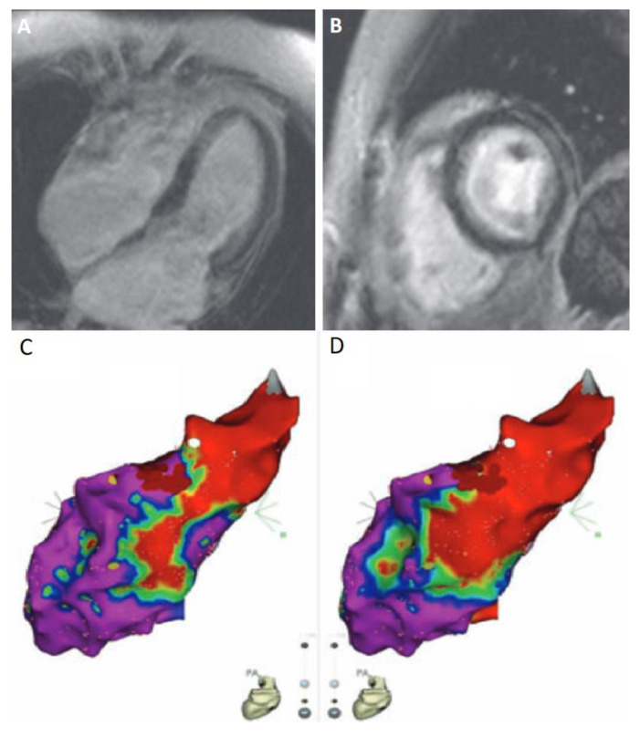
Figure 3.
Joint application of cardiac magnetic resonance (CMR) and electro-anatomical mapping (EAM) in the case of a 43 year old female diagnosed with Behçet’s disease and high burden of symptomatic ventricular extrasystoles (≈20,000 on 24 h ambulatory electrocardiography), without LV dysfunction. (A,B) T1-weighted sequence CMR with obvious LGE both at the left ventricular lateral wall (midmyocardial) and subendocardially (diffuse pattern, not corresponding to coronary artery territory). Four chamber (A) and short axis (B) views. (C,D) Electroanatomical map of the LV using the CARTO3® system (Biosense-Webster, Diamond Bar, CA, USA) depicting a low voltage area (non-purple color) in the same segments, using both bipolar (C) and unipolar (D) settings. Successful ablation of the arrhythmia was achieved following radiofrequency energy application at the basal lateral wall, an area with abnormal unipolar but not bipolar voltage values, suggesting a nonsubendocardial arrhythmogenic focus [116].

