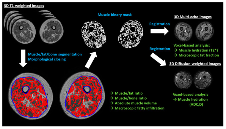
Figure 1.
Summary of the image analysis workflow. Muscle, fat, and bone are segmented from the T1-weighted images using differences in image intensities. The muscle binary mask is then closed to calculate the macroscopic fatty infiltration (relative volume of fat enclosed in the muscle volume, excluding bone volume). Binary volumetric masks of the muscle are spatially registered to the rest of MR images (multi-echo and diffusion-weighted), obtaining microscopic fat infiltration values (fat fraction) and hydration measurements (T2*, ADC (apparent diffusion coefficient) and D (diffusion)) on a voxel basis. White text indicates MRI sequences, blue indicates image analysis processes and green is for imaging biomarkers.

