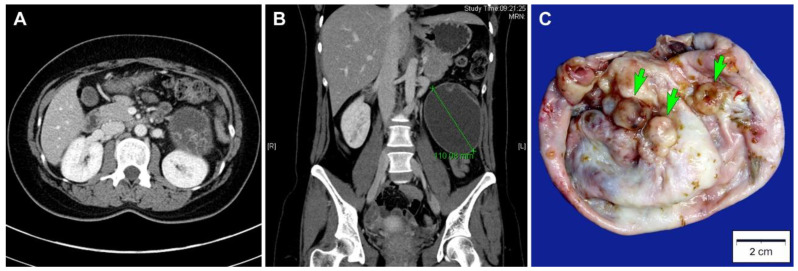
Figure 1.
Imaging and gross findings. (A) Abdominopelvic computed tomography scan in axial view, which revealed a thick-walled cystic mass in the left retroperitoneal space. The mass had some daughter cysts. (B) Abdominopelvic computed tomography scan in coronal view, which revealed a well-circumscribed, ovoid unilocular cystic mass with a diameter of 11 cm. (C) Grossly, the inner surface of the mass showed some round-to-ovoid, variegated mural nodules (green arrows), measuring up to 1.2 cm in the greatest dimension.

