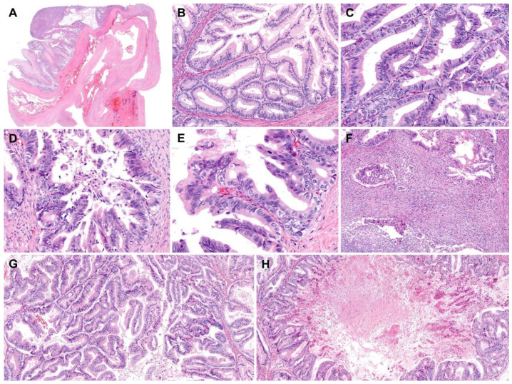
Figure 2.
Histological features of retroperitoneal mucinous carcinoma. (A) The thick fibrous wall showed mural hemorrhage. (B) Areas showing glandular proliferation were characterized by nuclear stratification and low-to-intermediate-grade nuclear atypia without stromal invasion, compatible with a mucinous borderline tumor. (C–E) In several foci, (C) high-grade nuclear atypia, (D) a micropapillary pattern, a loss of polarity, and (E) intraluminal papillary projections were noted. (F) Irregularly shaped cellular clusters and cribriform glands infiltrated the stroma, indicating microinvasion. There were associated stromal inflammatory infiltrates and desmoplastic reactions. (G,H) In addition to mucinous borderline tumors and microinvasive mucinous carcinomas, areas characterized by confluent glandular proliferation without intervening stroma were present, compatible with mucinous carcinomas with an expansile invasive pattern. A large, proliferating gland exhibited a cribriform architecture. Its lumen was extensively dilated and contained necrotic debris. Staining method: (A–H), hematoxylin and eosin. Original magnification: (A), 5×; (B), 40×; (C), 100×; (D), 100×; (E), 150×; (F), 40×; (G), 40×; (H), 20×.

