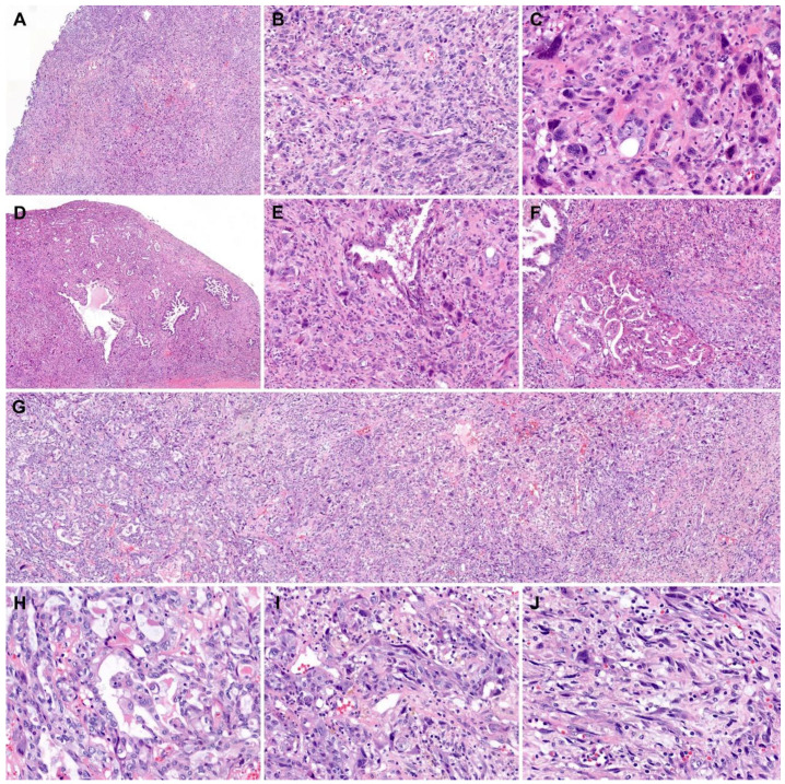
Figure 3.
Histological features of mural nodules associated with retroperitoneal mucinous carcinoma. (A) The mural nodules showed a diffuse proliferation of pleomorphic tumor cells, forming large, solid cellular sheets. (B) The spindle-shaped or polygonal tumor cells were arranged haphazardly. (C) Bizarre or multinucleated tumor cells were noted. (D) In some areas, variably sized, irregularly shaped glands were randomly distributed within the sarcomatous component. (E) An angulated tumor gland was embedded within the sarcomatous component. (F) Two large glands, which showed a complex, cribriform architecture, were present. (G–J) Sarcomatous transformation. The carcinomatous component displayed (H) a moderately differentiated adenocarcinoma (left one-third of image G) transformed through (I) a poorly differentiated carcinoma (middle one-third of image G) into (J) a sarcoma (right one-third of image G). Staining method: (A–J), hematoxylin and eosin. Original magnification: (A), 10×; (B), 100×; (C), 400×; (D), 10×; (E), 100×; (F,G) 40×; (H–J), 200×.

