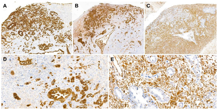
Figure 4.
Immunostaining results. (A,B) The carcinomatous component was highlighted using (A) cytokeratin 7 and (B) epithelial membrane antigens. (C) The sarcomatous component reacted with vimentin. (D,E) In high-power view, the carcinomatous and sarcomatous components showed mutually exclusive immunoreactivity to (D) pan-cytokeratin and (E) vimentin, respectively. Staining method: (A–E), polymer method. Original magnification: (A–C), 40×; (D,E), 200×.

