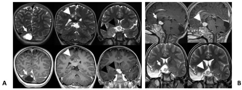
Figure 2.
(A) Coronal T2w (top row) and T1w (bottom row) images show cystic lesion in the cerebellum with a peripheral enhancing nodule (arrows), a small hyperintense lesion without contrast-enhancement involving the right gyrus cinguli (arrow heads). There is also a suprasellar/chiasmatic lesion (black arrows) with avid contrast-enhancement and cystic components, consistent with optic pathway glioma and a hyperintense and non enhancing lesion involving the right mesial temporal lobe (black arrow heads); (B) sagittal GdT1w (top row) and coronal T2w (bottom row) images show optic pathway glioma before (arrows) and during treatment (arrow heads).

