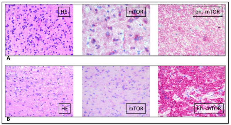
Figure 3.
(A) (upper raw) (low-grade glioneural tumor): HE: Low intensity glial proliferation with intermingled few dysplastic ganglion cells. Mild nuclear atypia and low proliferation index. mTOR: mild cytoplasmic positivity of neoplastic cells. Phospho–mTOR: strong cytoplasmic positivity of neoplastic cells; (B) (lower raw) (pilocytic astrocytoma): HE: Pilocytic glial proliferation in a fibrillary background with evidence of some Rosenthal fibers. Mild nuclear atypia and low proliferation index. mTOR: mild cytoplasmic positivity of neoplastic cells. Phospho–mTOR: Strong cytoplasmic positivity of neoplastic cells.

