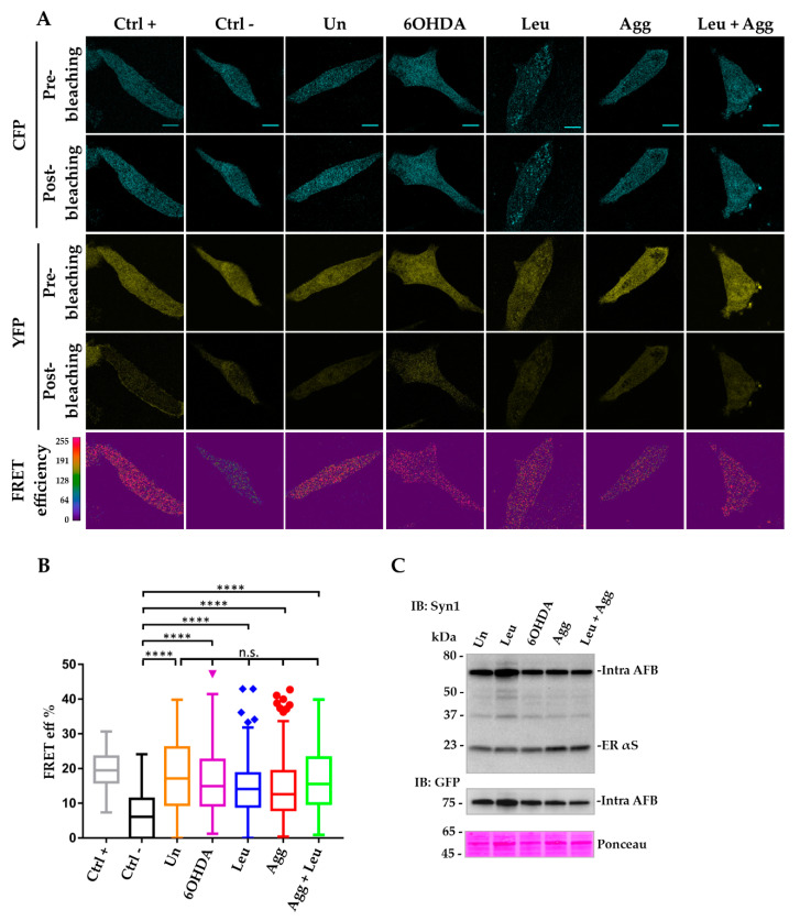
Figure 5.
FRET analysis of intra-AFB expressed in i36 shows that this reporter is sensitive to αS native structure but not to αS HMW species in pro-aggregating conditions. Induced i36 cells were transfected and assayed following the same protocol as described in Figure 4. (A) Images of AP FRET efficiency showed high FRET values (red regions) for the Ctrl + and for intra-AFB samples in untreated and treated conditions when compared to the Ctrl –. Scale bar, 10 μm. (B) Box Plot of FRET efficiency indicating that intra-AFB shows similar FRET values in untreated and treated conditions significantly different from the Ctrl –, but cannot distinguish between normal or stimulated states suggesting that intra-AFB is only suitable to sense native αS conformation. **** (p < 0.0001); n.s., not significant. (C) WB analysis of Tx-S fraction of cell lysates transfected with intra-AFB in untreated conditions or treated with pro-aggregating agents. Immunoblotting analysis with Syn-1 or GFP antibody of Tx-S fractions of i36 cell lines transfected with intra-AFB shows correct expression of the biosensor with the predicted molecular weight of about 75 kDa confirming the correct fusion of the two fluorescent probes to the same monomeric αS molecule.

