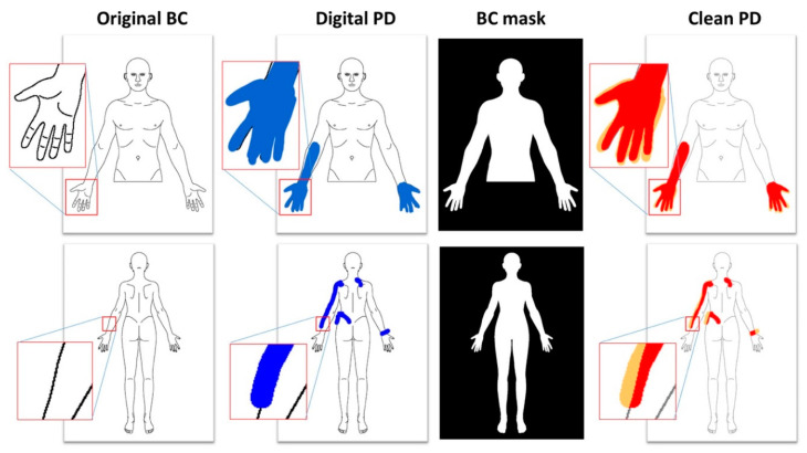
Figure 1.
Examples of identification of pain area from digital pain drawings. The original body chart is colored by the patient with a blue digital marker, then a mask is applied in order to remove the colored pixels outside the body chart area. The pixels of the clean pain draw (represented in red) are counted to obtain the pain area. The pixels outside the body chart area (represented in orange) are discarded.

