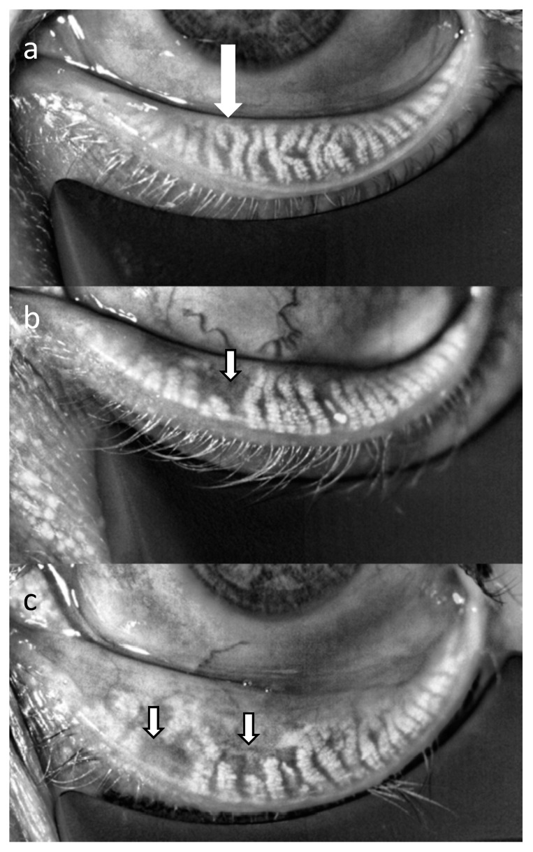
Figure 4.
Infrared images of the Meibomian glands of the lower eyelid in a healthy subject (a), a patient with mild Meibomian gland dysfunction (MGD) (b) and a patient with moderate MGD (c). Areas of gland dropouts can be clearly seen (arrows). The images were taken with the LipiView interferometer (TearScience, Morrisville, NC, USA).

