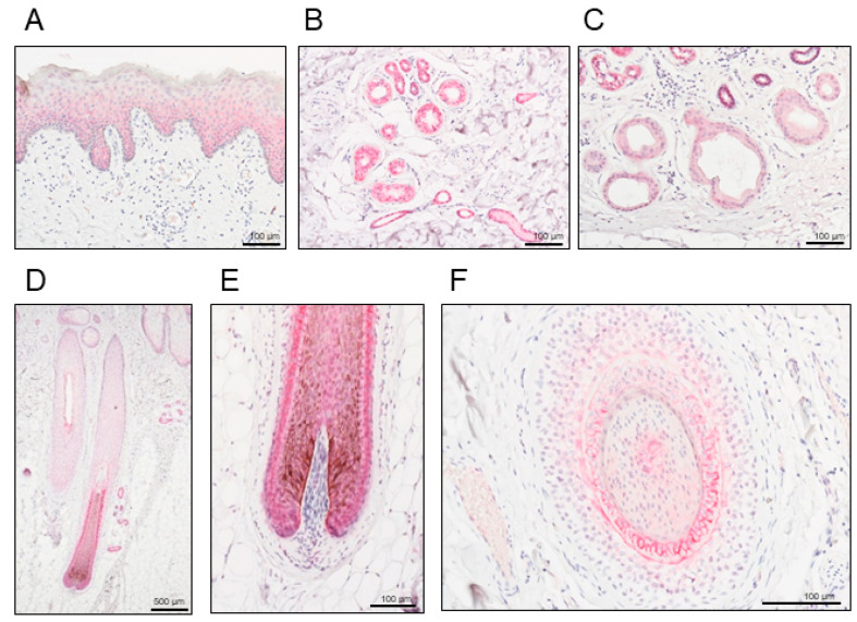
Figure 1.
Representative histopathological images of nectin cell adhesion molecule 4 (NECTIN4) staining in human normal skin. Positive signals are presented in red. (A) Epidermis, (B) eccrine sweat glands, (C) apocrine sweat glands, and (D–F) inner and outer root sheaths, and matrix of the hair follicles. Scale bars: 100–500 μm.

