
Fig. (4).
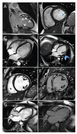
Cardiac magnetic resonance imaging findings in cardiac sarcoidosis. (A) Two-chamber view of LGE. (B) Four-chamber cine stack of lateral wall thinning. (C) Uniform wall on the first scan. (D) Four-chamber view of wall thinning. (E) and (F) SA stack of LGE. (G) and (H) Three-chamber view of LGE. LGE, late gadolinium enhancement. (A higher resolution / colour version of this figure is available in the electronic copy of the article).