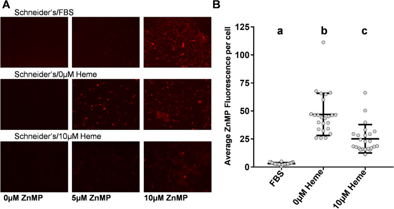
Fig. 1.
Heme deficiency in Aag2 cells increases ZnMP uptake. a Images of ZnMP fluorescence in cells incubated in various heme concentrations. Brightness and contrast were increased uniformly in all images by reducing the pixel range from 0 to 255 to 0–150. b Quantitative measurement of 5 μM ZnMP normalized fluorescence of cells exposed to heme treatments. Each dot represents the average normalized ZnMP fluorescence of a well of a 96-well plate after 30-min incubation. The middle horizontal line on each treatment group represents the mean and the lines above and below represent ±1 SD. Each heme treatment was distinct from each other (q-val < 0.01) and thus labeled a, b &c

