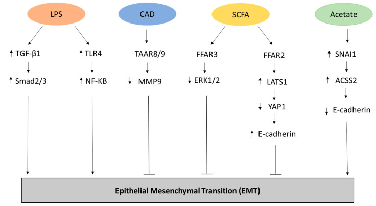
Figure 1.
Representation of the metabolites and related pathways involved in the Epithelial-Mesenchymal transition (EMT) process in cancer. Lipopolysaccharide (LPS) has been shown to promote EMT through the upregulation of transforming growth factor beta-1 (TGFβ-1) and Mothers Against Decapentaplegic Homolog 2/3 (Smad2/3) [22] as well through the increased expression of Toll Like Receptor 4 (TLR4) and NF-KB [23] in biliary epithelial cells. Conversely, Cadaverine (CAD) can inhibit EMT in breast cancer cell lines through the activation of trace amino acid receptors 8 and 9 (TAAR8/9) modulating the expression of metalloproteinase 9 (MMP9) [29]. In breast cancer cell lines, inhibition of EMT can be carried out through the metabolic pathway of short chain fatty acids (SCFAs) that activates Free Fatty Acid Receptor 2 (FFAR2), leading to inhibition of the Hippo-Yap pathway and increased expression of adhesion protein E-cadherin, and FFAR3, resulting in mitogen-activated protein kinase (MAPK) signaling inhibition [30]. Finally, acetate can promote EMT by increasing the expression of the zinc finger protein Snail Family Transcriptional Repressor 1 (SNAI1) and Acyl-CoA Synthetase Short Chain Family Member 2 (ACSS2) in renal carcinoma cells under glucose limitation [31].

