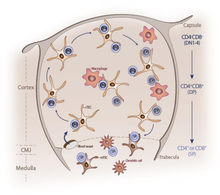
Figure 1.
Schematic representation of post-natal thymus. The outer mesenchymal capsule enters the thymus at regular intervals to form trabeculae. Inside, the developing thymocytes are embedded in a three-dimensional network of thymic stroma, mainly composed of thymic epithelial cells (TECs), dendritic cells, endothelial cells, macrophages, and fibroblasts. Each thymus compartment delimited by trabeculae is divided into two histologically distinct regions, the cortex and medulla, separated by the corticomedullary junction (CMJ). Immature hematopoietic progenitors enter the thymus via the vasculature at the CMJ and commit to the T-cell fate. Thymocytes migrate from the CMJ to the subcapsular zone of the cortex, as they differentiate through CD4–CD8– double-negative 1-4 (DN1-4) stages to the CD4+CD8+ double-positive (DP) stage. DP cells interact with cortical TEC (cTEC), differentiate into either CD4+ or CD8+ single-positive (SP) cells, and migrate back to the CMJ. DP cells positively selected to mature into CD4+ or CD8+ single positive (SP) cells then cross the CMJ and enter the medulla, where they undergo the final stages of maturation before being exported to the periphery. Self-reactive SP cells are deleted by ‘negative selection’, mediated by thymic dendritic cells and medullary TECs (mTEC).

