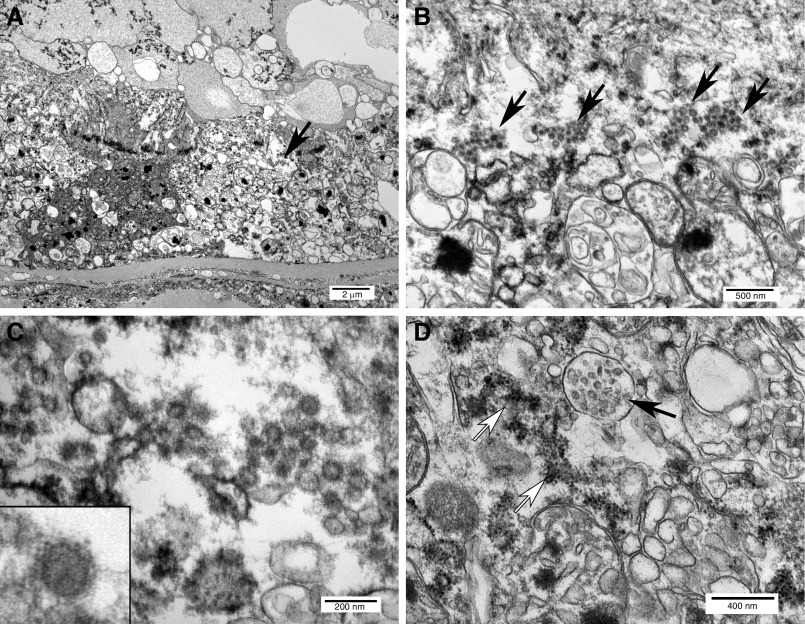
Figure 3.
Ultrastructural features of coronavirus infection and replication in proximal tubular epithelial cells detected after death from COVID-19. (A) Proximal tubule oriented with basement membrane at the bottom and lumen at the top, containing vacuolated and partially degenerated epithelial cells with abundant viral particles (arrow). (B) Intracytoplasmic viral arrays (arrows) within tubular epithelial cells. (C) Detail of viruses showing envelope with crown-like projections. Inset: single virus. (D) Vacuole with double-membrane vesicles (solid arrow) adjacent to ribosome-studded rough endoplasmic reticulum (open arrows), similar to structures reported in SARS-CoV-1–infected cells.

