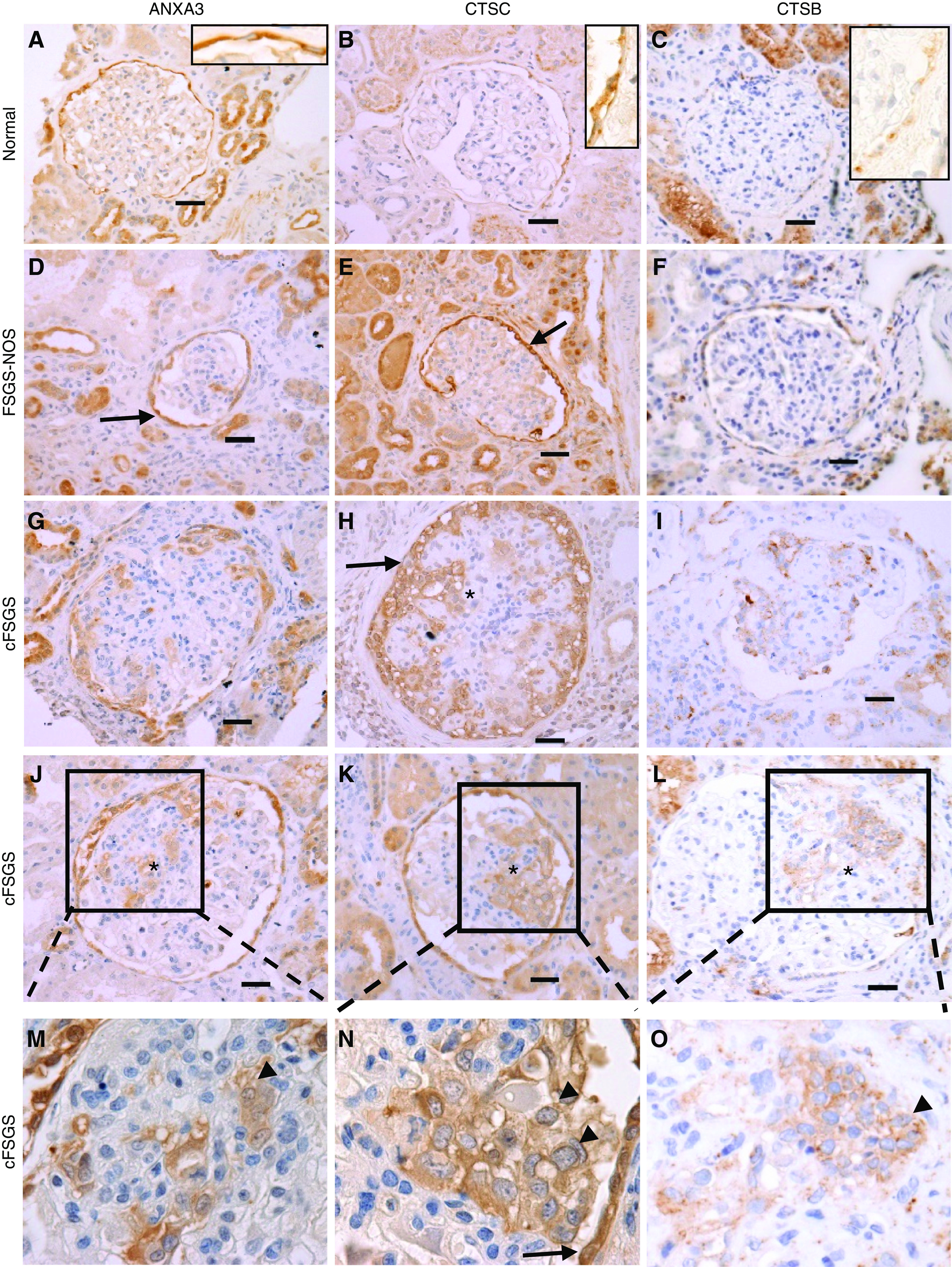
Figure 2.

Glomerular tufts stain for annexin A3, cathepsin B, and cathepsin C in cFSGS. Glomerular expression of annexin A3, cathepsin C, and cathepsin B was visualized by IHC. Representative photomicrographs of glomeruli are shown for protein staining in (A–C) NCs, (D–F) FSGS-NOS, and (G–O) cFSGS. Arrows identify PECs along Bowman’s capsule showing hypertrophy and enlarged nuclei. (M–O) Show enlargements of the boxes in (J–L) showing infiltration of glomerular tufts by stained cells (arrowheads). The asterisk marks areas of glomerular tuft staining. Scale bars, 30 μm. Original magnification, 40×.
