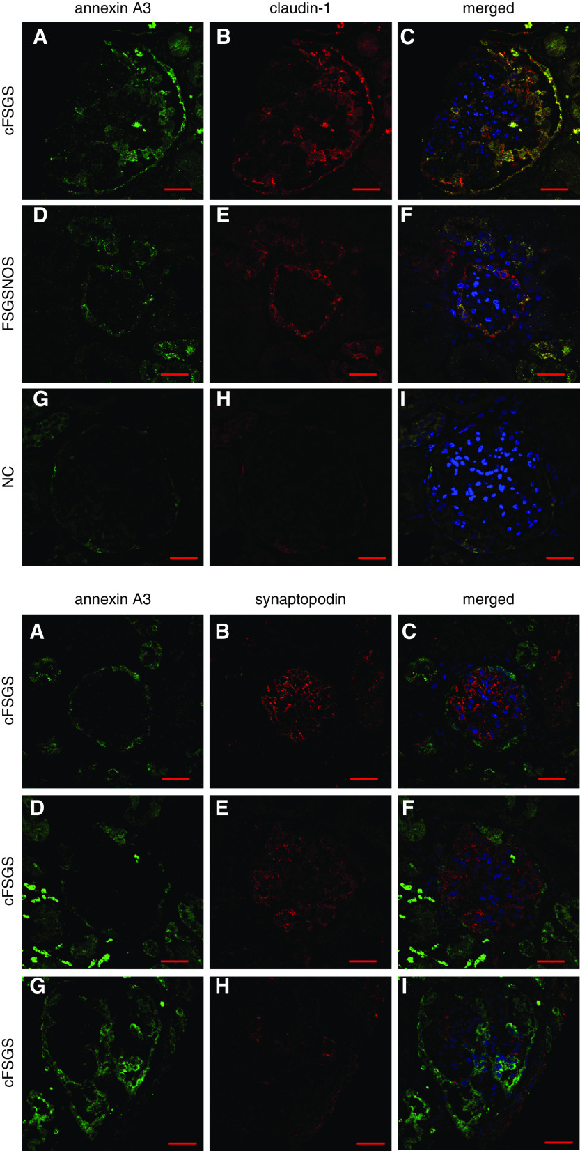
Figure 4.
Annexin A3 colocalizes with claudin-1, but not synaptopodin in cFSGS. (A) Sections of renal biopsies from patients with (A–C) cFSGS, (D–F) FSGS-NOS, and (G–I) NCs immunostained for annexin A3, claudin-1, and nuclei stained with 4′,6-diamidino-2-phenylindole. The panels show staining for (A, D, and G) annexin A3 only, for (B, E, and H) claudin-1 only, and (C, F, and I) merged images. The merged images show that annexin A3 and claudin-1 colocalize. Photomicrographs are 1-µm, single-plane images acquired by confocal microscopy. Original magnification of all images, 60×. Scale bars, 30 µm. (B) Sections of a renal biopsy from a patient with cFSGS immunostained for annexin A3, synaptopodin, and nuclei stained with 4′,6-diamidino-2-phenylindole. The panels show staining for (A, D, and G) annexin A3 only, for (B, E, and H) synaptopodin only, and (C, F, and I) merged images, in three different glomeruli from the same patient. The merged images show that annexin A3 and synaptopodin do not colocalize. Photomicrographs are 1-µm, single-plane images acquired by confocal microscopy. Original magnification of all images, 60×. Scale bars, 30 µm.

