
Figure 5.
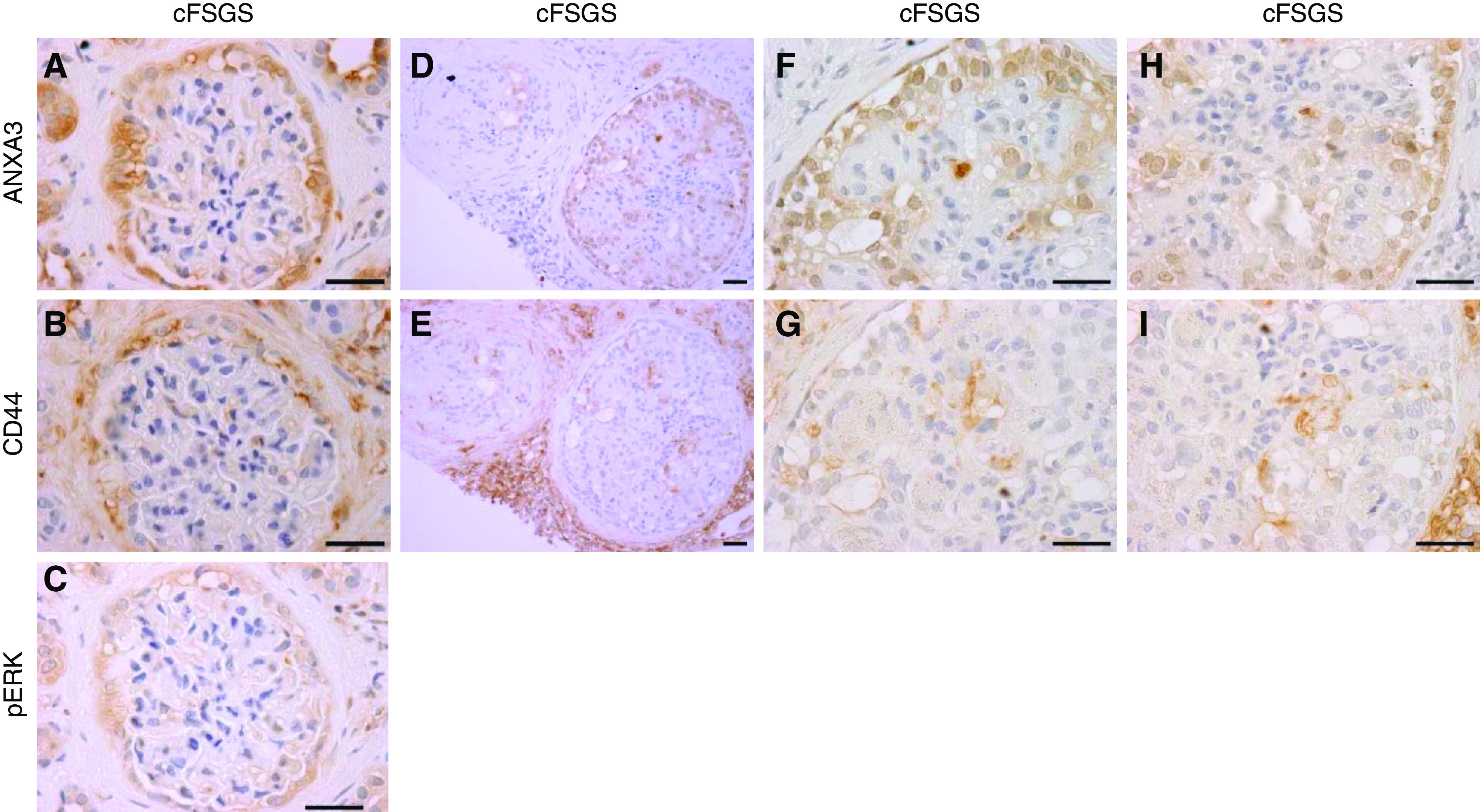
Annexin A3–stained PECs express CD44 and pERK in cFSGS. Photomicrographs show IHC staining patterns for (A) annexin A3, (B) CD44, and (C) pERK in serial sections of a cFSGS glomerulus. PECs showed hypertrophy/hyperplasia, enlarged nuclei, and adhesions to glomerular tufts. Annexin A3, CD44, and pERK showed the same staining pattern. (D and E) Serial sections of a single cFSGS glomerulus with annexin A3–stained PECs infiltrating the glomerular tuft, demonstrating CD44 staining of plasma membranes (E). (F–I) Enlarged portions of the same glomerulus as in (D and E). Scale bars, 30 µm. Original magnification, 100× for (A–C) and (F–I), and 40× for (D and E).