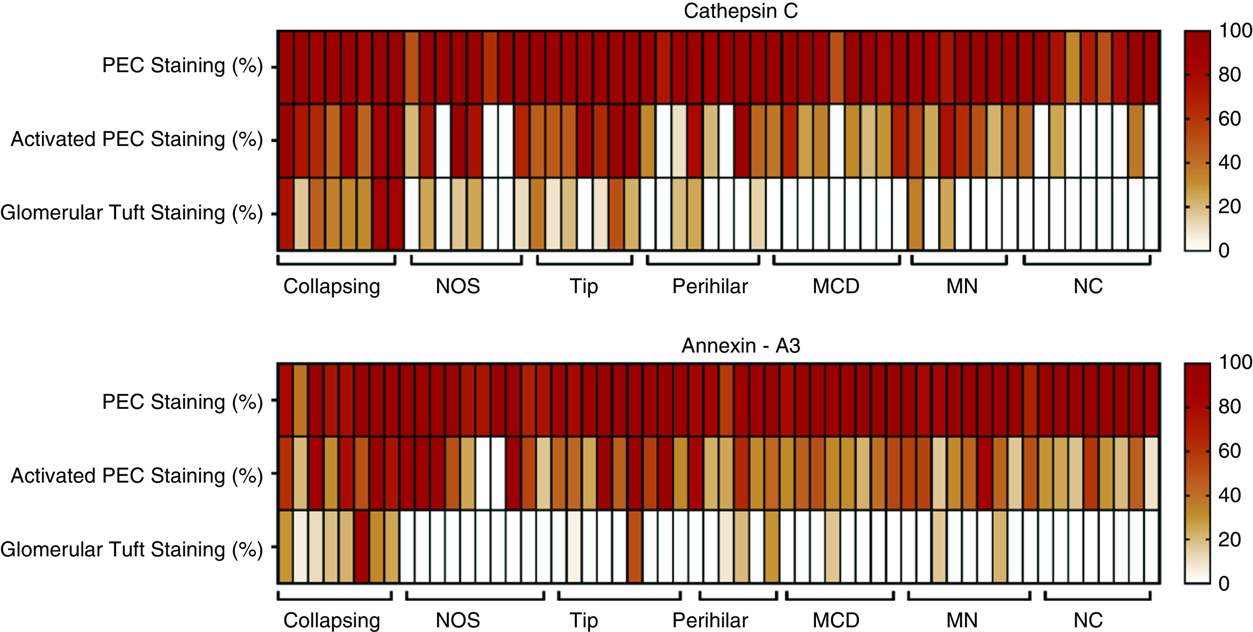
Figure 6.

Enhanced annexin A3 and cathepsin C staining in glomerular tufts in cFSGS, compared to other proteinuric glomerular diseases. Heat maps show the percentage of glomeruli with annexin A3 and cathepsin C staining of PECs with normal or hyperplastic morphology and cells infiltrating glomerular tufts for all patients with FSGS variants, MCD, MN, or no glomerular disease (NC).
