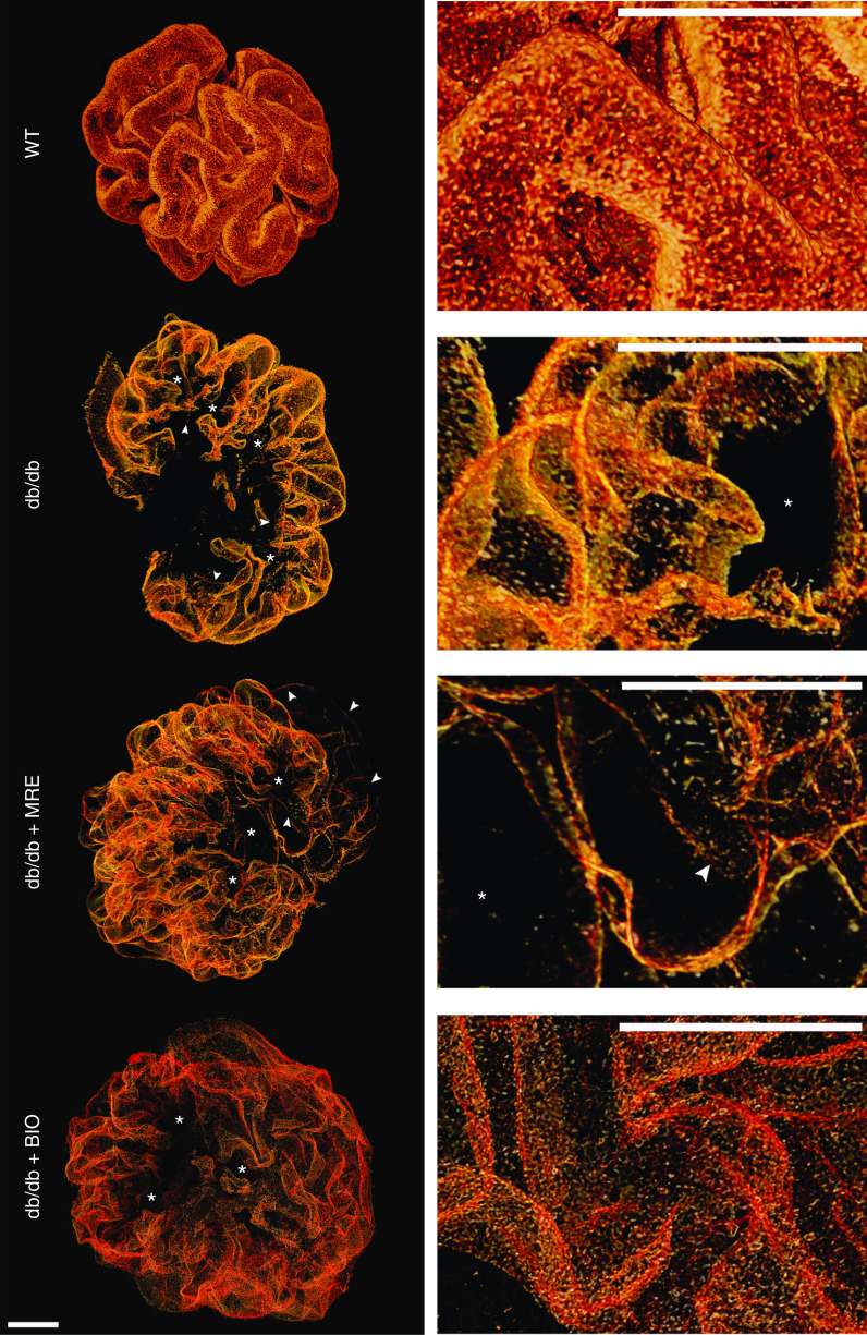
Figure 4.
Three-dimensional reconstruction of glomeruli stained for nephrin upon optical tissue clearing. Images show representative glomeruli of mice from each group. Signal represents nephrin protein within the slit diaphragm. Z-series stacks were obtained from 80-μm kidney slices. Images were collected at 1-μm intervals. WT 1K mice show smaller glomeruli with an intact foot process coverage of the glomerular capillaries, i.e., the intact glomerular filtration barrier. Db/db 1K mice show large defects in coverage representing denudated areas (black areas) with podocyte loss (marked with an asterisk). Arrowheads indicate filtration slits between podocytes showing foot process effacement. MRE and BIO treatment reduced the area of denudation, and BIO especially reduced signs of foot process effacement. Bars, 20 µm. Magnification, ×40.

