Figure 4.

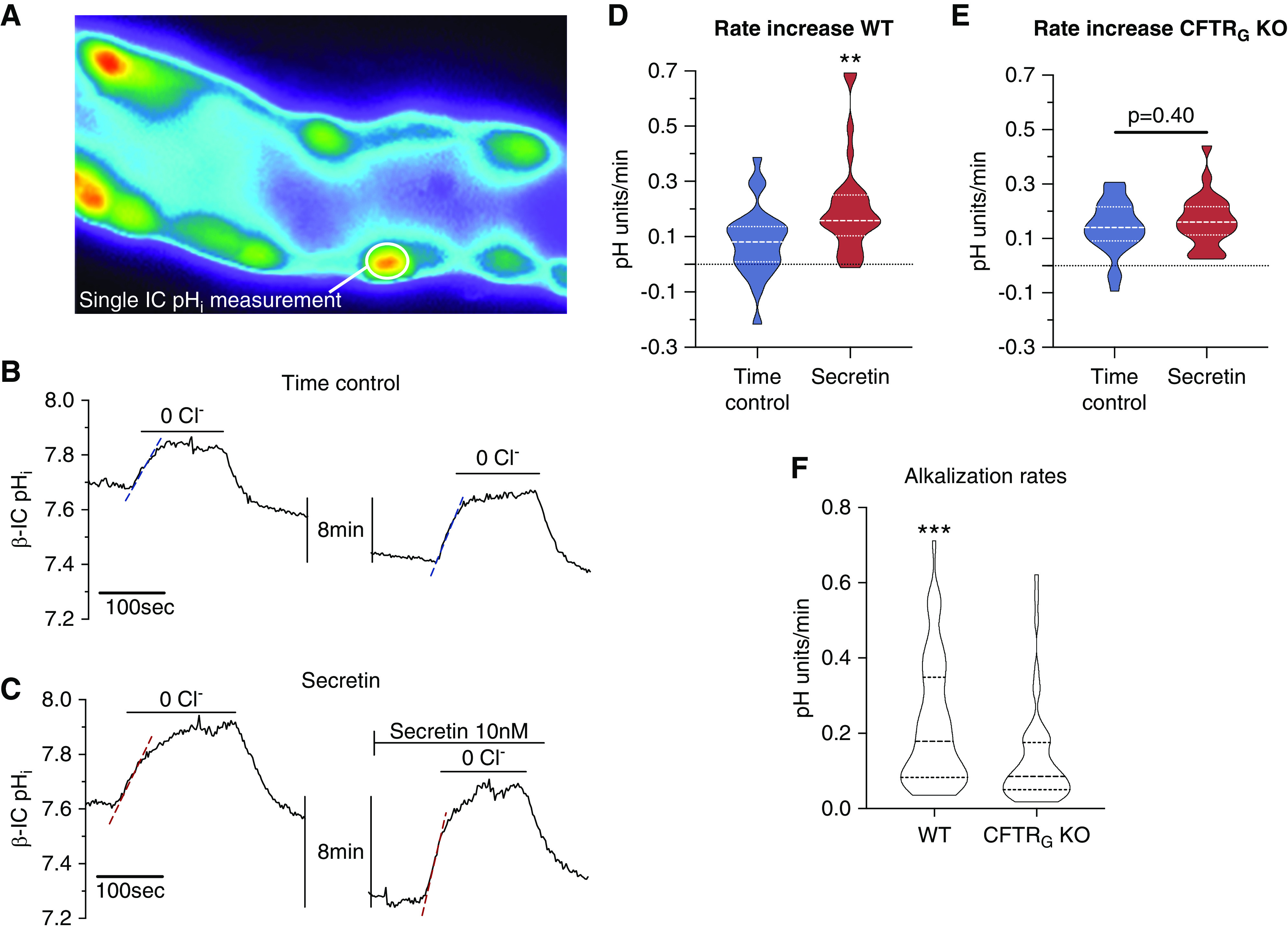
Secretin stimulates pendrin-mediated HCO3− transport in isolated mouse CCD. (A) Fluorescence image of an isolated perfused CCD loaded with luminal BCECF-AM. Multiple intercalated cells protruding into the lumen can be identified. Most of the intercalated cells in the CCD show functional pendrin positivity (prompt intracellular alkalization after luminal chloride removal, data not shown). (B) A time control experiment showing an original pHi recording of a single pendrin-positive cell after a first removal of luminal chloride followed by a second removal of luminal chloride 10 minutes later. (C) A secretin stimulation experiment showing an original pHi recording of a single pendrin-positive cell. Luminal chloride was removed twice, first without stimulation and second after stimulation with 10 nM basolateral secretin for 10 minutes. Note the markedly faster alkalization effect in this experiment after secretin stimulation. (D and E) Summary of the entire experimental series. Secretin stimulated a significantly faster intracellular alkalization rate as compared with time controls, indicating activation of pendrin-mediated HCO3− secretin (n=5 tubules, n=23–28 cells, t test). (F) Note the significantly lower baseline functional pendrin activity in CFTRG KO CCDs (n=10 tubules, n=51–53 cells, t test). **P<0.01, ***P<0.001.
