
Figure 1.
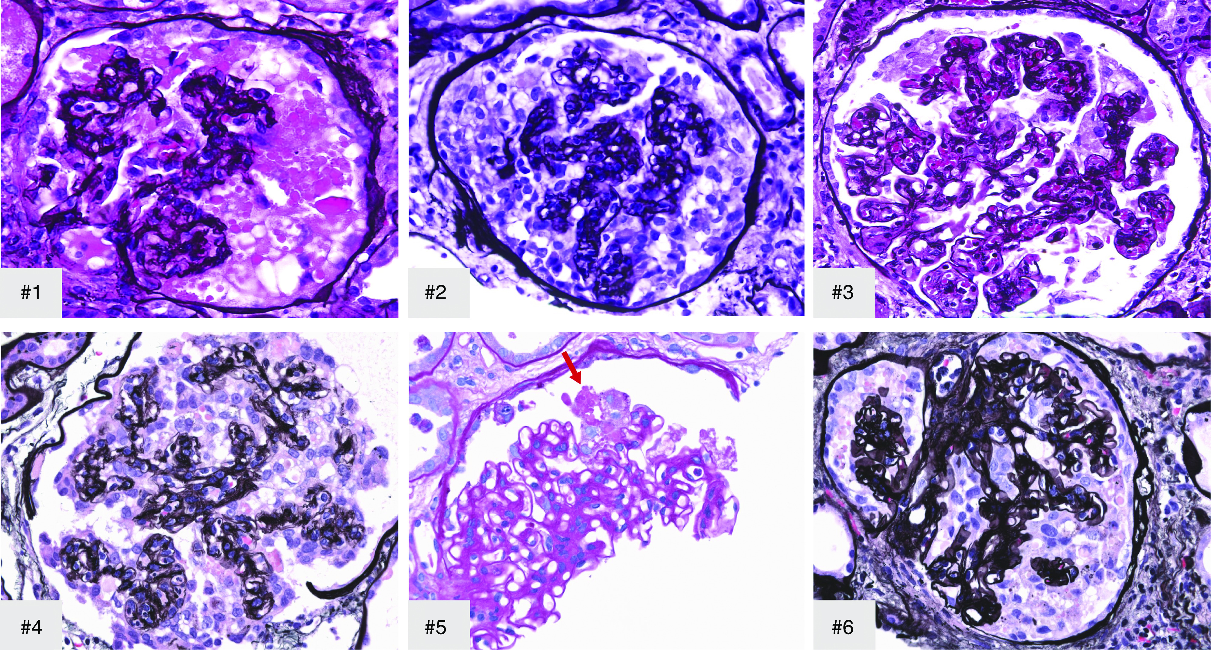
Collapsing glomerulopathy, showing collapse of glomerular tuft with overlying hypertrophy and hyperplasia of visceral epithelial cells and marked protein droplets in cases 1–6. Arrow, early segmental collapsing lesion. #1–#4 and #6, Jones methenamine silver; #5, Periodic acid–Schiff; original magnification, ×500 in #1 and ×400 in #2–#6.