
Figure 2.
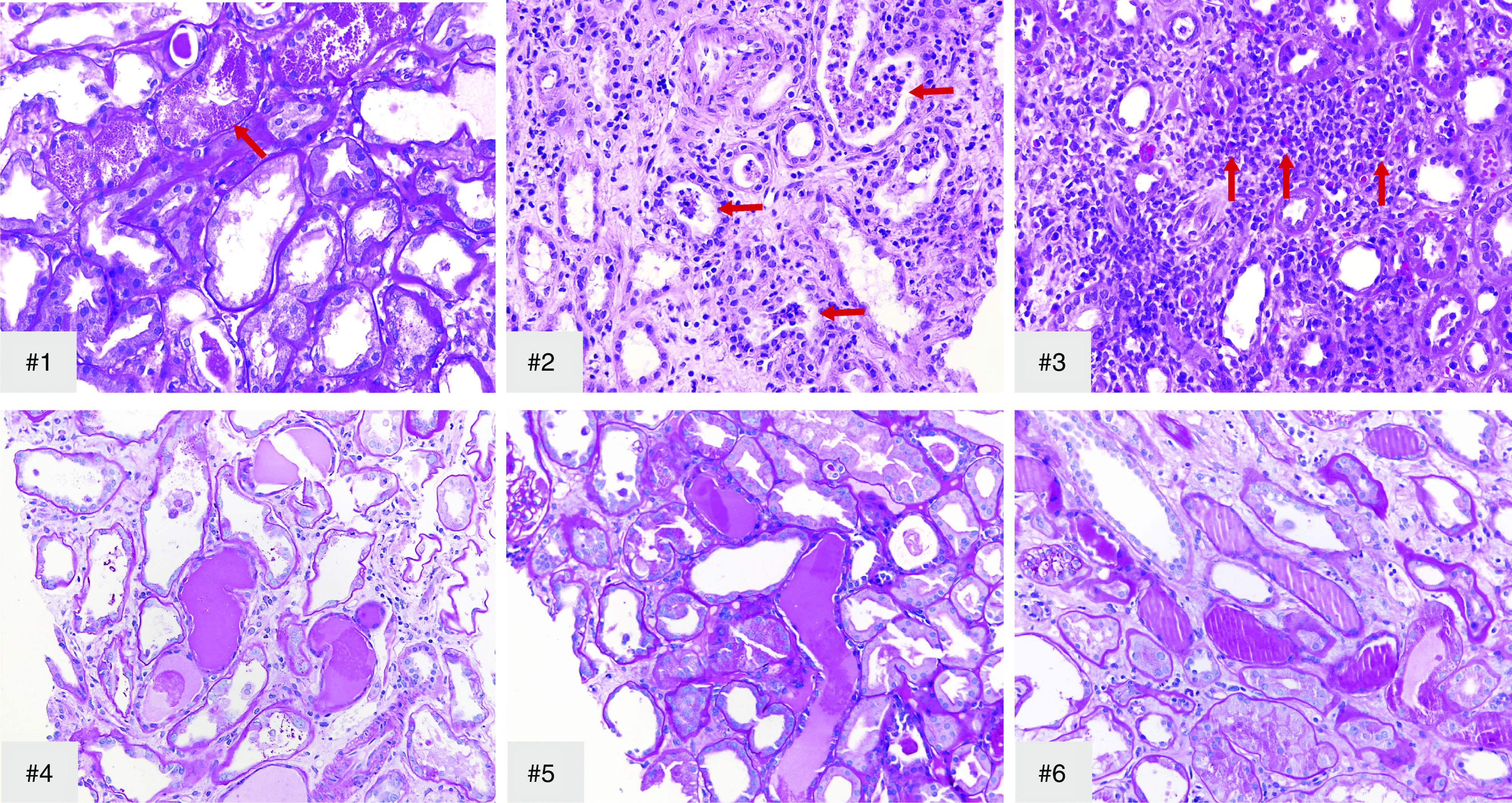
All biopsies show acute tubular injury with frequent tubular protein droplets. Arrow in #1, tubular protein droplets. In addition, case 2 (#2) showed diffuse patchy pleomorphic interstitial infiltrate with neutrophils and tubulitis, and rare tubules with intratubular neutrophils (arrows); case 3 (#3) showed focal acute interstitial nephritis with occasional foci of eosinophils (arrows), and microcystic tubules were present in all cases except #3. #1, #4–#6, Periodic acid–Schiff; #2 and #3, hematoxylin and eosin; original magnification, ×400 in #1–#3 and ×200 in #4–#6.