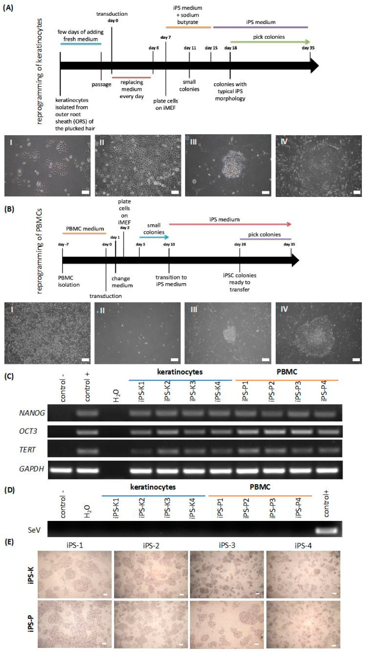
Figure 1.
Reprogramming of keratinocytes and peripheral blood mononuclear cells (PBMCs) to induced pluripotent stem cells (iPSCs) with Sendai viral vector. Characterization involved 4 clones of each origin of iPS cells. (A) Scheme presenting generation of iPS cells from plucked hair keratinocytes. (AI) Representative image of the cells four days after transduction. (AII) Morphology of the tightly-packed cells, which is characteristic of the early stage of reprogramming. (AIII) Formation of the first colonies on day 14 after transduction. (AIV) A big and tightly packed colony, which is ready to be transferred, between 18 to 35 days. White scale bars represent 200 μm. (B) Scheme presenting generation of iPS cells from PBMCs. (BI) Representative image of PBMCs in the expansion medium five days before transduction. (BII) PBMCs one day after transduction with Sendai viral vector. (BIII) Formation of small colonies on day 16. (BIV) Big and tightly packed colony, ready to be transferred on day 28. White scale bars represent 200 μm. (C) Keratinocyte-derived-iPS (iPS-K) and PBMC-derived-iPS (iPS-P) cells express endogenous pluripotency genes, such as NANOG, OCT3 and TERT. Positive control (control+) was a commercially available protein-induced iPS cell line (piPS), whereas negative control (control−) was a human colon cancer cell line, HTC116. (D) Viral transgene (SeV) was silenced in the generated cell lines after the 10th passage. Positive control for Sendai virus genome transgene was the generated iPS cell line at an early passage, whereas negative control was the piPS cell line. (E) Generated iPS clones displayed alkaline phosphatase activity. White scale bars represent 400 μm. Pictures show representative images. NANOG—homeobox protein NANOG; OCT3—octamer binding transcription factor 3; TERT—telomerase; GAPDH—housekeeping gene, glyceraldehyde 3-phosphate dehydrogenase; SeV—Sendai Virus genome.

