-
A
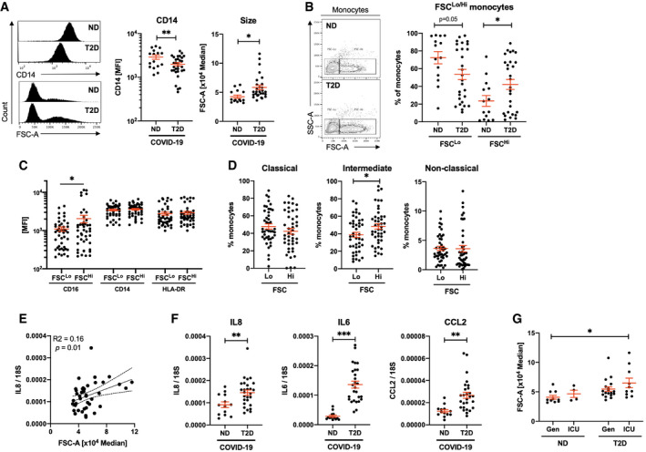
Quantification of CD14 expression and size (FSC) in monocyte populations from non‐diabetic (ND) and type 2 diabetic (T2D) COVID‐19 patients.
-
B
Frequency of conventional (FSCLo) and large (FSCHi) monocytes in ND and T2D COVID‐19 patients.
-
C
Expression of CD16, CD14 and HLA‐DR in FSCLo and FSCHi monocytes from COVID‐19 patients.
-
D
Proportions of classical, intermediate and non‐classical monocytes within FSCLo and FSCHi monocytes from COVID‐19 patients.
-
E
Correlative analyses of IL8 expression in peripheral blood mononuclear cells (PBMCs) to monocyte FSC.
-
F
IL8, IL6 and CCL2 mRNA expression in PBMCs from ND and T2D COVID‐19 patients.
-
G
Monocyte size quantified in ND and T2D COVID‐19 patients treated in general wards (Gen) or in the intensive care unit (ICU).
Data information: Data are presented as mean ± SEM. Differences between groups were evaluated with unpaired
t‐test (except for (C) and (D) where paired by patient). Analysis of variance (ANOVA) or analysis of covariance (ANCOVA) was used for multiple group comparisons. *
p < 0.05; **
p < 0.01 and ***p < 0.001. For correlative analysis, Spearman's test was carried out calculating a 2‐tailed
P‐value. See also
Appendix Fig S2. Sample size and exact
P‐value in
Appendix Table S5.
Source data are available online for this figure.