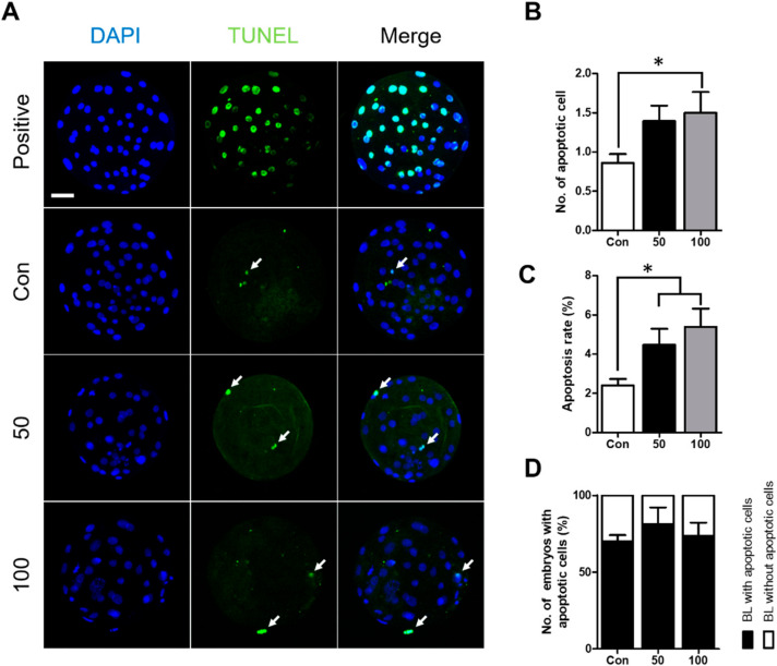
Figure 2.
Effect of TCS exposure on the survival of blastomeres in parthenogenetically activated porcine embryos. (A) Detection of apoptosis in blastocysts cultured with different concentrations of TCS. Bar = 50 μm. The Blue DAPI signal (left), green terminal deoxynucleotidyl transferase-mediated dUTP-digoxygenin nick end-labeling (TUNEL) staining (middle), and a merged image (right) are shown. White arrows indicate TUNEL-positive cells. (B) Number of apoptotic blastomeres in blastocysts. (C) Proportion of apoptotic blastomeres in blastocysts cultured with different concentrations of TCS. (D) Quantification of proportion of blastocysts with and without apoptotic cells. Data are from three independent experiments, and the values represent means ± SEM (* p < 0.05).

