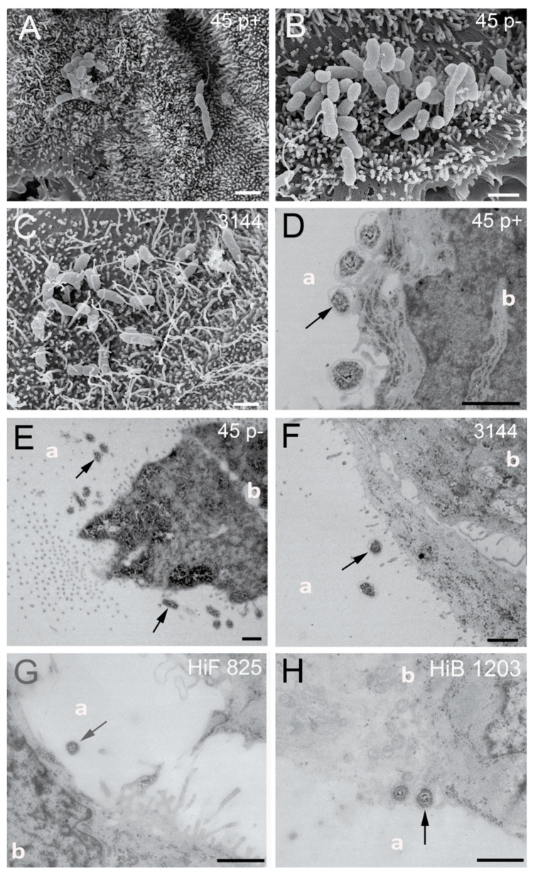
Figure 6.
SEM and TEM sections of HIBCPP cells grown in the standard cell culture system infected with indicated NTHI and H. influenzae isolates for 6 h: (A–C) SEM images of NTHI strains 45 p+, 45 p− and 3144, which are located at the microvilli covered apical cell side; and (D–H) TEM images of bacteria adhering to the cells sitting on or next to microvilli of the HIBCPP (black arrows). Almost no intracellular bacteria could be detected in the standard culture grown HIBCPP cells. The apical and basolateral cell sides are labeled with “a“ and “b”, respectively. Scale bars represent 1 µm.

