Fig. 4.
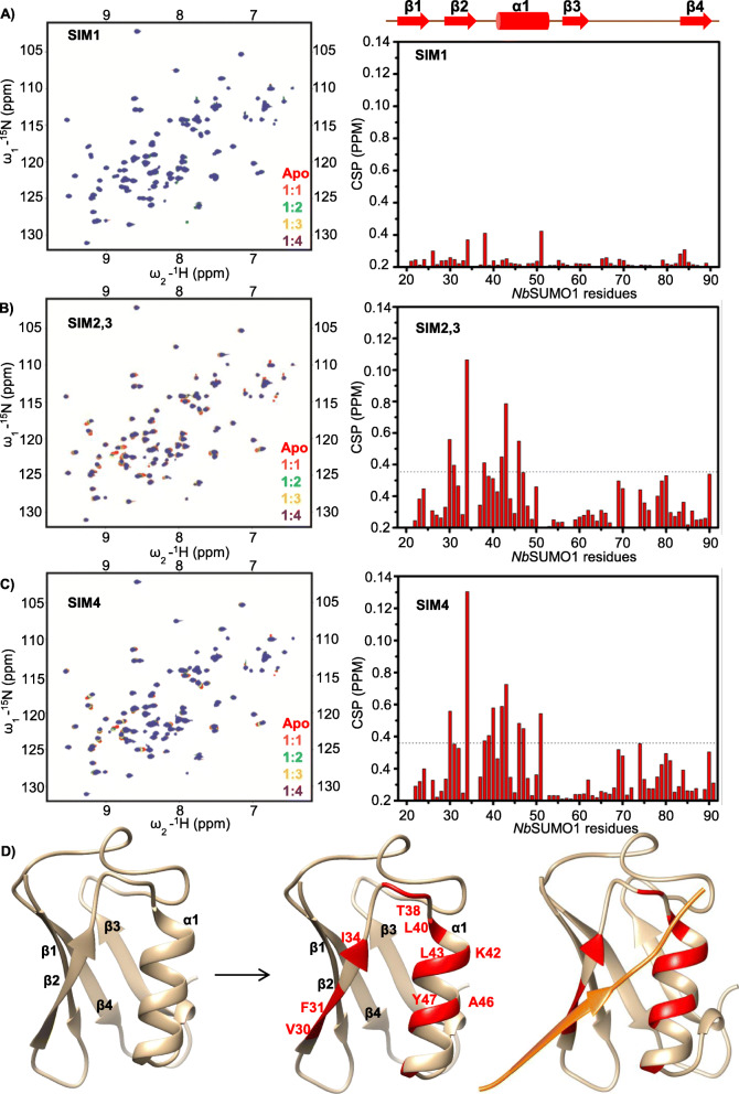
βC1 SIMs interact with NbSUMO1 as seen with 15N-1H HSQC spectrum. a SIM1, b SIM2,3 and c SIM4 (left panel). The right panel shows corresponding CSPs between free and NbSUMO1-SIM-bound form plotted against individual residues of NbSUMO1. The dashed line indicates mean ± SD of CSP values. Residues above the dash line show binding interface. d Structure of NbSUMO1. Left panel: NbSUMO1 predicted structure, ribbon and surface representation. Middle panel: residues of NbSUMO1 interacting with βC1 SIM motifs (Red). Left panel: NbSUMO1/βC1-SIM4 model. SIM4 is shown in orange

