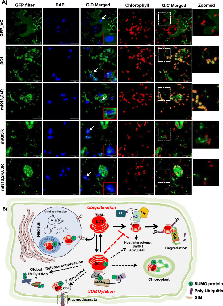
Fig. 7.
Mutations in SUMOylation motif of βC1 alters its localization. a GFP-tagged βC1 and its SUMOylation motif mutants were introduced onto N. benthamiana leaves as described in the “Methods” section. The nucleus was stained with DAPI, and chlorophyll auto-fluorescence was measured at 650 nm. βC1 localization in the nucleus and nucleolus is shown with a white arrow and a representative nucleus was zoomed in the red box. The white box with dashed line represents the zoomed area from G/C merged panel. G/D merged, GFP and DAP filter merged; G/C, GFP and chlorophyll auto-fluorescence merged. Size bar 50 μM. b Schematic representation showing interplay of multiple PTMs regulating function of βC1. Host ubiquitination machinery recognizes βC1 and an unknown ubiquitin ligase ubiquitinates βC1 via interaction with its SIM motif leading to its degradation. This inhibits viral movement. Viral counter-defence is achieved by interaction of βC1 with host SUMOylation machinery. SUMOylated βC1 is stable, and this aids in the movement of the virus and pathogenicity

