Fig. 4.
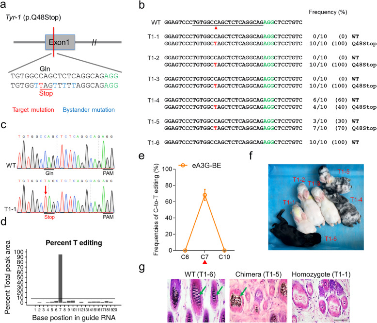
Generation of Tyr p.Q48stop rabbits mimic human OCA1 using eA3G-BE system. a Target sequence at the Tyr-1 (p.Q48stop) locus. PAM region (green), target mutation (red), and bystander mutation (blue). b Alignments of mutant sequences of F0 rabbits from T-A cloning. The targeted sequence is underlined. The PAM site and base conversions are shown in green and red, respectively. The column on the right indicates frequencies of mutant alleles. WT, wild-type. T1–1 to T1–6, each individual. c Representative sequencing chromatograms from a WT and mutant rabbit (T1–1). The red arrow indicates the substituted nucleotide. The relevant codon identities at the target site are presented under the DNA sequence. d The predicted editing bar plot based on Sanger sequencing chromatograms from T1–1 by EditR. e Summary of single C-to-T editing frequencies of F0 rabbits at Tyr-1 using eA3G-BE. f Photograph of all six F0 rabbits at 1 week. g H&E staining of skin from WT (T1–6) and Tyr-1 mutant (T1–1 and T1–5) rabbits. The green arrows highlight the melanin in the basal layer of the epidermis. Scale bars 50 μm

