
FIGURE 1.
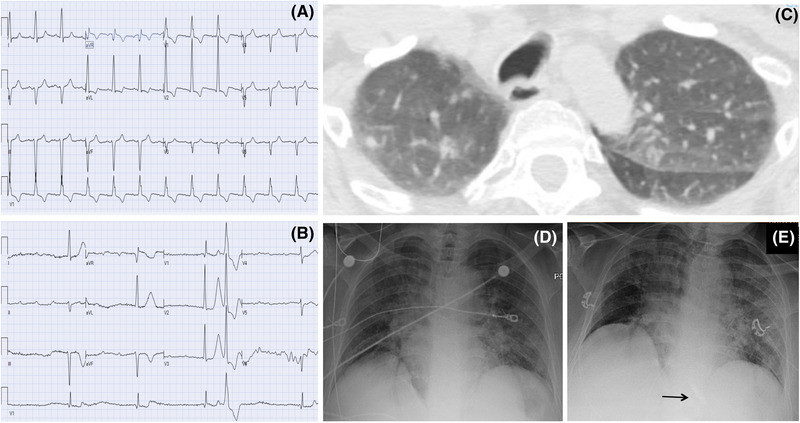
A patient presenting with AV block and imaging findings consistent with COVID‐19 prior to development of pulmonary symptoms. A, Baseline ECG prior to admission. B, ECG on presentation demonstrating third‐degree AV block. C, CT on admission with incidental findings of ground‐glass nodular opacities in the lung apices. D and E, Chest X‐ray from admission, and following leadless pacemaker implant (arrow) showing progressive bilateral pulmonary infiltrates [Color figure can be viewed at wileyonlinelibrary.com]