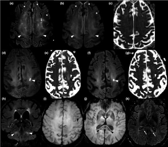
Figure 2.

Brain magnetic resonance imaging (MRI) findings in the five patients studied. Case 1: Axial fluid‐attenuated inversion recovery (FLAIR)‐weighted MRI (a), axial diffusion‐weighted MRI (b) and axial apparent diffusion coefficient (ADC) map (c). MRI images obtained on day 21; cross‐sections through motor areas, frontal and parietal lobes. Diffuse bilateral, symmetric white matter FLAIR hyperintensities with mild hyperintense 'ground glass' areas (arrow heads) and frank hyperintense areas (conventional radiological FLAIR hyperintensities; arrows). All abnormal FLAIR areas appeared hyperintense in the diffusion sequence and were characterized by a gradient: frank hyperintense FLAIR lesions had a higher intensity than 'ground glass' areas. On the ADC map, 'ground glass' areas were iso‐ or hypointense, whereas frank hyperintense FLAIR areas were hyperintense. Abnormal FLAIR hyperintensities were preferentially localized to subcortical white matter of motor areas and showed a bilateral symmetric distribution. The anterior frontal white matter appeared normal on FLAIR and diffusion‐weighted imaging (DWI) sequences (a, b: stars). Case 2: Axial DWI (d) and axial ADC map (e): cross‐sections through frontal and parietal lobes on day 17. Axial DWI (f) and axial ADC map (g): cross‐sections through frontal and parietal lobes on D33. Coronal FLAIR weighted MRI on day 33 (h): cross‐section through middle cerebellar peduncles. The first MRI (on D17) revealed an acute hyperintense DWI lesion, with hypointensity on ADC map, suggestive of a cytotoxic edema in the left frontal lobe (d, e) and deemed initially compatible with an acute stroke. However, this lesion maintained a similar aspect following 16 days (f, g), casting doubts on its ischaemic origin. Persistence of middle cerebellar peduncles hyperintensities was also evident (h). Case 4: Axial susceptibility‐weighted imaging MRI at day 31: cross‐section through motor areas, frontal and parietal lobes (i) and the splenium (j). Axial FLAIR weighted MRI (k): cross‐section through the splenium. Multifocal microbleeds were evident in the splenium (j: black arrow heads) and in the white matter/gray matter junction (i: black arrow head), with an apparent perivascular distribution in the Virchow‐Robin spaces. These lesions were associated with hyperintensities on FLAIR weighted sequence (k: arrows). Day 0 = date of COVID‐19 symptom onset.
