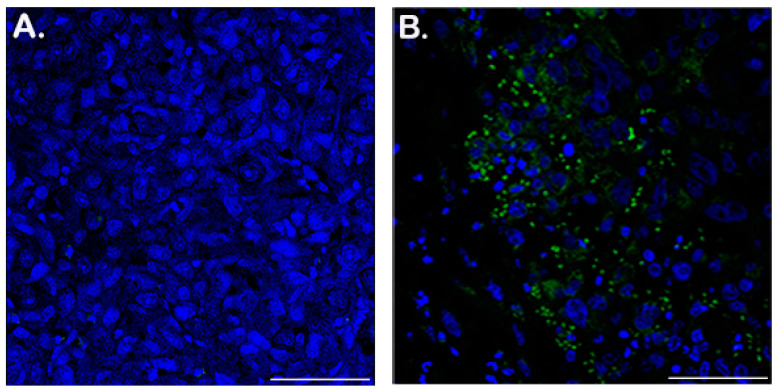
Figure 3.
Immunohistochemistry of PC3-LgBiT tumor sections show HiBiT-reporter virus persistence within tumor xenografts 43 days post infection. Virus infected cells assessed via hexon detection in tumor xenografts. Frozen sections of tumors injected with PBS (A) or HiBiT-reporter virus (B) were stained by immunohistochemistry for adenovirus detection with an anti-hexon antibody and an Alexa Fluor 488-labeled secondary antibody; the sections were counterstained with Hoechst in TBS. (A) Representative control section from a tumor injected with PBS. (B) A representative section from virus-infected tumors at 43 days post infection. Size of the scale bar is 100 µm.

