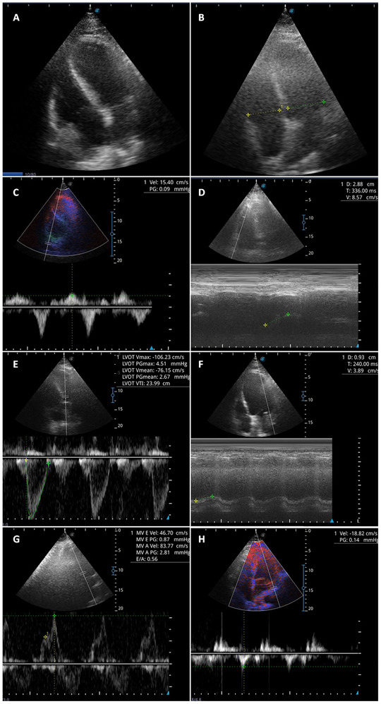
FIGURE 2.

Evaluation of right and left ventricular function in mechanically ventilated patients in prone position. Panel A: Apical 4‐chamber view. Panel B: right ventricle/left ventricle ratio. Panel C: tricuspid peak systolic S wave tissue Doppler velocity (S wave).Panel D: tricuspid annular plane systolic excursion (TAPSE). Panel E: left ventricular outflow tract velocity integral time (LVOT VTI). Panel F: mitral annular plane systolic excursion (MAPSE). Panel G: E velocity of mitral inflow filling pattern. Panel H: lateral e′ velocity
