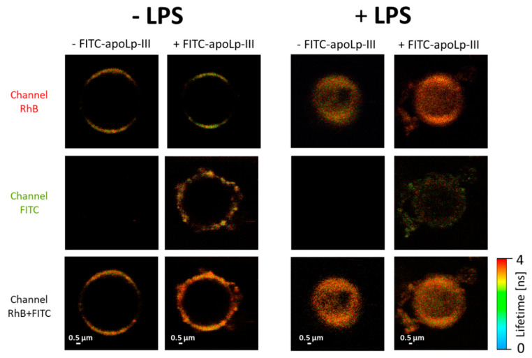
Figure 7.
FLIM images of giant unilamellar vesicles (GUVs) formed with DPPC with (+LPS) or without (−LPS) L. dumoffii LPS. The vesicles were exposed (+apoLp-III) or not exposed (−apoLp-III) to FITC-apoLp-III. The upper panel shows the fluorescence emission recorded in the channel representing Rhodamine B (RhB) present in the lipid phase and the middle panel shows fluorescence emission recorded in the channel representing FITC (Figure S2 for a specification of the fluorescence emission channels). The lower panel shows superposition of fluorescence signals recorded in both channels.

