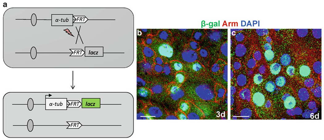
Fig. 3.

Tubulin-lacZ positive-labeling to generate ISCs clones in the midgut. (a) Diagram to show the lineage-tracing scheme to mark the randomly dividing cells by heat shock FLP-catalyzed site-specific recombination. (b, c) The representative examples of the induced clones, 3 day (b) and 6 day (c) in the midgut anti-β-Gal (green), anti-arm (red), and Dapi (blue). Scale bars: 10 μm.
