
Figure 3.
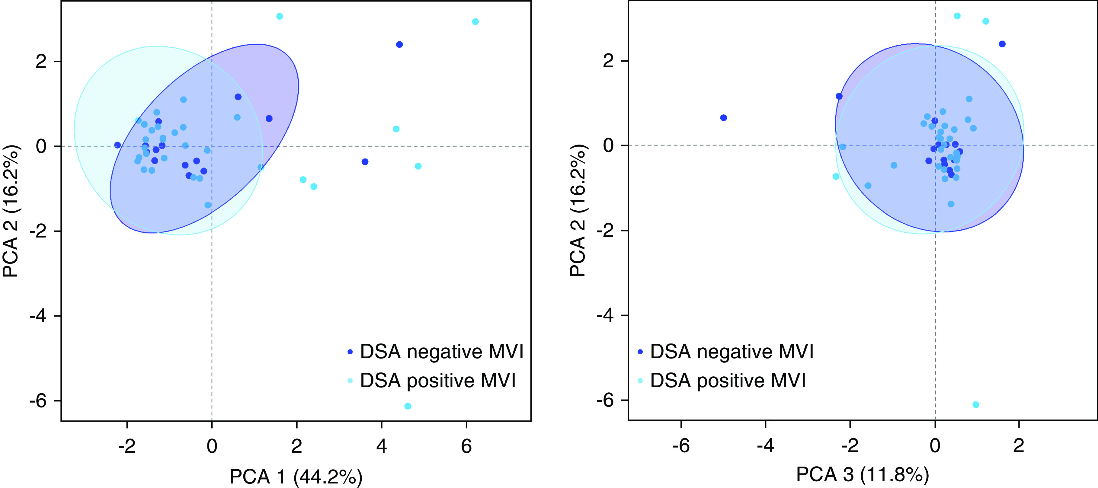
Spatial distribution and extent of leukocyte infiltration in MVI is not influenced by HLA-DSA. Principal component analysis (PCA) of leukocyte subset infiltration quantified by computer-assisted analysis of immunohistochemical staining in kidney allografts with MVI, in the presence (n=32) or absence (n=15) of circulating HLA-DSA. Plotted 95% confidence ellipses show major areas of overlap.