
Figure 1. Optimization of detection of human P-2 (hP-2) mRNA by FISH-Flow RNA Assay.
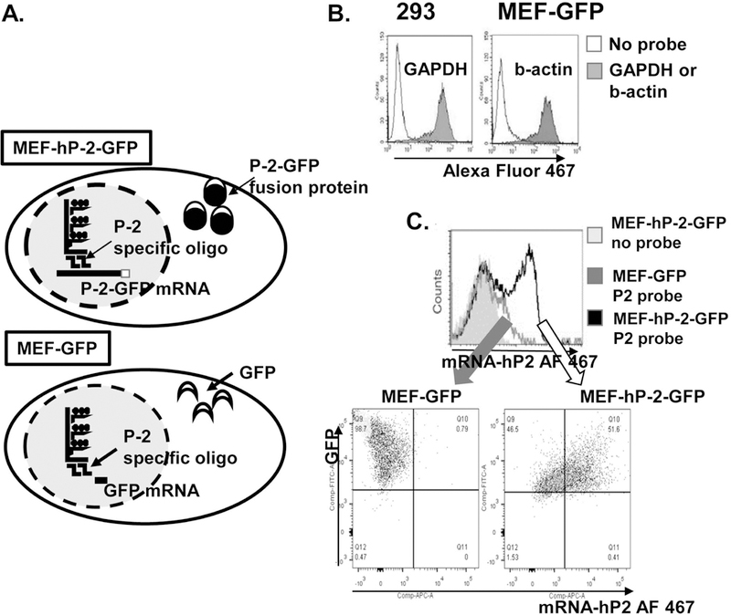
a. Human P-2 gene can be easily detected in a mouse embryonal fibroblast (MEF) cell line overexpressing human P-2-GFP fusion protein used as a positive control for the FISH-Flow Assay optimization. b. Flow cytometry data showing glyceraldehyde 3-phosphate dehydrogenase (GAPDH) RNA expression in 293 cells and mouse b-actin in MEF cell line expressing GFP used as negative controls for the assay optimization. c. Representative overlay histogram showing hP-2 mRNA and gfp protein expression in MEF-GFP and MEF-hP2GFP cells by flow cytometry. The signal-to-noise ratio was calculated as the ratio between the mean fluorescence intensity (MFI) of the hP-2 gene and the MFI of the no probe control.