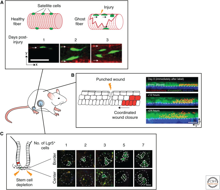
Figure 4.
Following stem cells during regenerative processes by intravital microscopy. (A) Intravital microscopy reveals the dynamics of satellite cell-mediated repair of injured muscle fibers. Remnants of damaged fibers form ghost fibers, which are used by satellite cells as guides for proliferation and migration. Scale bar, 50 µm. (Photos in panel A are reprinted from Webster et al. 2016, with permission, from Elsevier © 2016.) (B) Imaging of the cell dynamics during wound healing in the skin reveals the migratory and proliferative dynamics of the cells repairing the wound (migratory and proliferative cells are shown in red). Scale bar, 50 µm. (Photos in panel B are reprinted from Park et al. 2017, with permission, from Springer Nature © 2017.) (C) Multiday intravital microscopy shows the highly plastic nature of the intestinal cells. After ablation of all the stem cells, transit-amplifying cells fall back into the stem cell niche, adopt a stem cell fate, and repopulate the stem cell zone. Repopulating transit-amplifying cells are depicted in green. Scale bar, 20 µm. (Photos in panel C are adapted from Ritsma et al. 2014, with permission, from the authors.)

