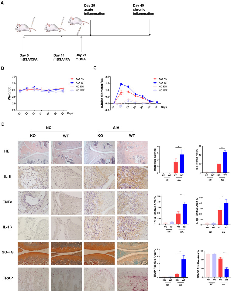
FIGURE 3.
AdipoR1 knockout in T cells ameliorates antigen-induced arthritis (AIA) disease. (A) Timeline of AIA induction. (B) Weights were recorded daily after the 3rd immunization (n = 5). (C) Mean diameters of knee joints were recorded daily after the 3rd immunization (n = 5). (D) Photomicrographs for knee joint sections stained with H&E on day 25 (original magnification 100×) (n = 3–5). Photomicrographs of immunohistochemical staining of IL-6, TNFα and IL-1β in knee joint tissues on day 25, positive cells were stained with intense brown color (original magnification 400×) (n = 3). Knee joints were stained for TRAP positivity (left) on day 25 post AIA (original magnification 400×) and quantified by computer analysis (n = 3). Proteoglycan staining was demonstrated by safranin O-fast green immunostaining (original magnification 200×) (n = 3) (*p < 0.05, **p < 0.01, and ***p < 0.001).

