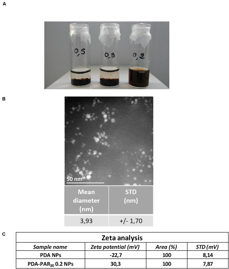
FIGURE 2.
(A) Picture showing the stability after 2 months at 4°C of the different PDA-PAR30 NPs formulations using different concentrations of dopamine hydrochloride (0.5, 0.3, and 0.2 mg.mL–1) with a defined concentration of polyarginine (PAR) of 1 mg.mL–1. (B) Typical image of PDA-PAR30 0.2 NPs obtained with Transmission Electron Microscope (TEM) and the subsequent size quantification results. (C) Results of zeta potential measurements performed on PDA-PAR30 0.2 NPs as compared to pure PDA particles.

