Figure 1.
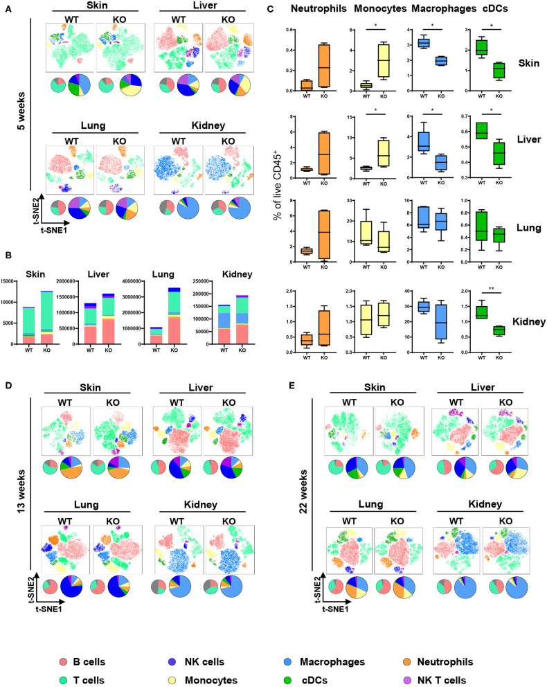
Altered immune cell populations upon partial PTPN2 deletion in DCs. Immune cells were analyzed in 5-weeks-old, 13-weeks-old, and 22-weeks-old PTPN2fl/fl (WT) and PTPN2fl/fl × CD11cCre (KO) mice. t-SNE maps displaying live, CD45+ single cells in skin, liver, lung, and kidney. Colors correspond to FlowSOM-guided clustering of cell populations. Pie charts represent relative numbers among CD45+ cells. (A) t-SNE maps of CD45+ cells in 5-weeks-old mice. (B) Total counts of CD45+ cells among indicated tissues in 5-weeks-old-mice. (C) Relative numbers of neutrophils, monocytes, macrophages, and DCs among CD45+ cells among indicated tissues in 5-weeks-old mice. (D,E) t-SNE maps of CD45+ cells in 13-weeks-old (D) and 22-weeks-old (E) mice. Data are representative of two independent experiments with n ≥ 4 mice (A–E). *p < 0.05; **p < 0.01 [two-tailed Mann Whitney test (C)]. Data are shown as mean ± s.d. (C).

