FIG. 9.
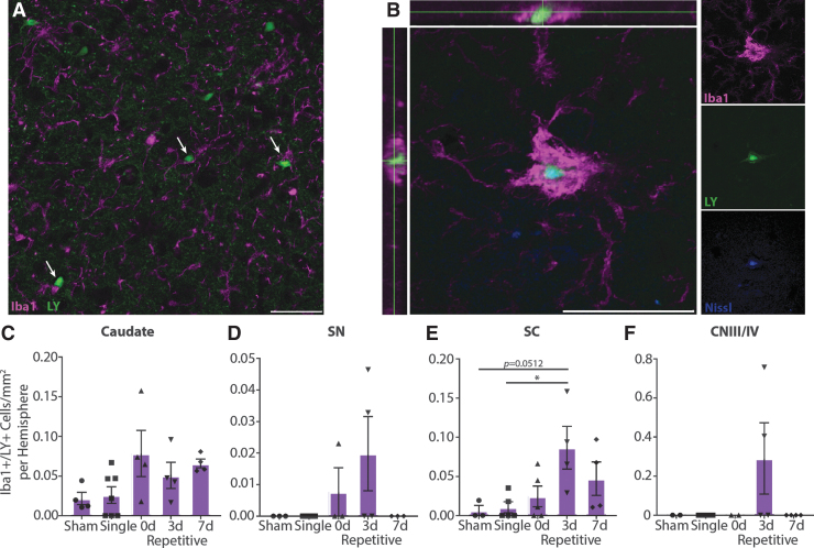
Microglia interacting with permeabilized neurons. (A) In repetitively injured animals, microglia often contact Lucifer Yellow (LY)+ neurons (arrows). Representative image from caudate of 3-day (3d) repetitive injury. (B) Large microglia can be observed colocalized with LY+/Nissl+ cells, suggesting phagocytosis of permeabilized neurons. Representative image from caudate of 7-day (7d) repetitive injury. Iba1, purple; LY, green; Nissl, blue. Scale bars = 50 μm. (C-F) Quantification of large Iba1+ signal colocalized with LY, ± SEM. Phagocytosis occurs more frequently in repetitively injured animals, especially those injured 3 days apart, although these trends were only statistically significant in the superior colliculus (SC; caudate p = 0.0680; substantia nigra pars reticulata [SNpr] p = 0.1457; SC p = 0.0273; cranial nerve III/IV [CNIII/IV] p = 0.2000). Color image is available online.

