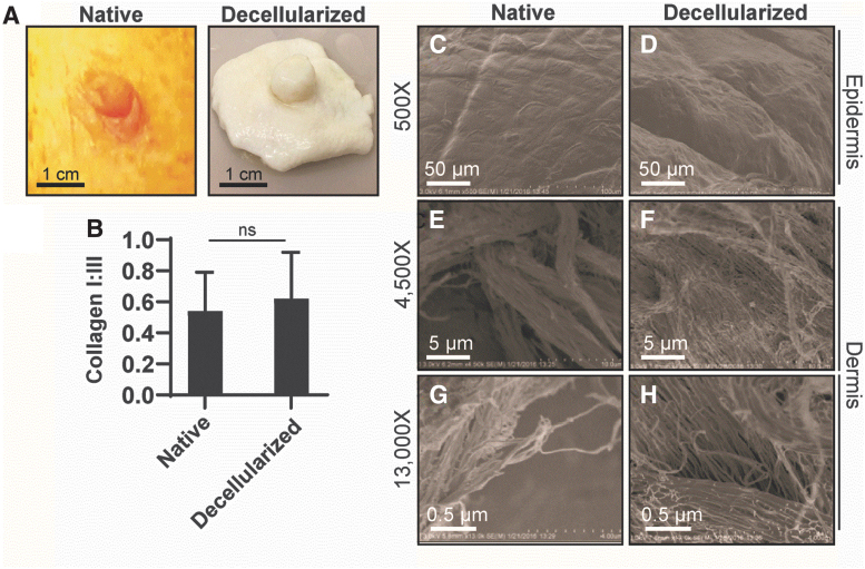
FIG. 4.
Structure of the rhesus macaque nipple. (A) Photographs of an NHP-NAC (native) and dcl-NHP-NAC (decellularized). (B) Graph of collagen I:III levels in native and decellularized NACs. Student's t-test. ns, nonsignificant (p > 0.05). (C–H) Scanning electron cryomicroscopy images of native and decellularized NACs. (C, D) Micrographs showing the epidermis. (E–H) Micrographs showing the dermis at different magnification.

