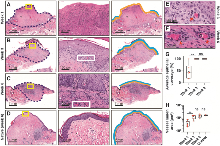
FIG. 7.
NHP-mediated recellularization of decellularized nipple grafts. Examples of NHP-nipple grafts resected after (A) 1 week postengraftment, (B) 3 weeks postengraftment, and (C) 6 weeks postengraftment. (D) The native NHP-nipple after 6 weeks postengraftment. The second column shows magnified regions of the yellow boxes in the first column of (A–D); and the third column indicates how epithelialization coverage was calculated: yellow lines are the entire exposed area of the grafts and blue lines are the reepithelialized region of the exposed grafts. (E, F) Magnified regions from white boxes in (B, C) showing examples of neovascularization (red arrows). (G) Graph of average epithelial coverage of nipple grafts over time. (H) Graph of blood vessel lumen area from nipple grafts over time. The control sample is of the host's native nipples from 6 weeks postengraftment. One-way ANOVA with Tukey's post hoc test was performed in (G, H). **p < 0.01. ns, not significant.

